Multimodal digital training system for neurosurgery
The spectrum of everyday operations in neurosurgery includes a large number of different operations and surgical techniques. Depending on the size and catchment area of the respective neurosurgery clinic, certain surgical procedures are performed less than 5-10 times a year, meaning that a learning curve can rarely occur. At the same time, as in all surgical disciplines, patients’ expectations of neurosurgeons have increased significantly. They rightly expect a surgeon with the appropriate experience for each procedure. Resection of the tumor with a safety margin is not possible on the brain. Every brain tumor operation is associated with a high level of technical and personnel expenditure. Neuronavigation, microscope, ultrasound and various image modalities must be understood and coordinated for effective interpretation and use. This can only be learned through standardized and objectifiable training of the neurosurgeon for all aspects of tumor surgery and cannot be achieved solely in practice on the patient in the operating room. A training system for learning, understanding and training the procedures involved in brain surgery is therefore urgently needed.
Together with the Department of Neurosurgery at Leipzig University Hospital, PHACON is currently developing a multimodal digital training system for neurosurgery.
This project is financially supported by funds from the European Union and the Free State of Saxony.
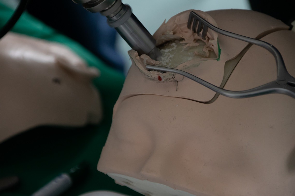
The aim of the project is to develop a neurosurgical trainer with which it is possible to process patient-specific data sets and thus map different tumor localizations for the surgeon. Such an approach enables a flexible and broad range of applications. The surgeon goes through all stages. Starting with the planning using their own software, through to the execution on the realistic brain model and the evaluation of the procedure. A new approach is added to the procedure on the model. Using an augmented reality scenario, additional content and images are projected directly into the surgeon’s field of vision via glasses. This innovative approach offers the advantage of combining the real world of the haptic phantom with a virtual world that becomes visible through the glasses. This enables training with real instruments and haptic feedback, while additional virtual content such as pathologies and risk structures can also be displayed